Draw A Labelled Diagram Of A Eukaryotic Plant Cell As Seen In An Electron Micrograph : Topic 1 2 Cell Ultrastructures Ppt Download : In truth, there are still features of plant and animal cells we're only lately here is an electron micrograph of an animal cell with the labels superimposed:
byJoshua Albrecht-
0
Draw A Labelled Diagram Of A Eukaryotic Plant Cell As Seen In An Electron Micrograph : Topic 1 2 Cell Ultrastructures Ppt Download : In truth, there are still features of plant and animal cells we're only lately here is an electron micrograph of an animal cell with the labels superimposed:. The nucleus is an organelle present in the cytoplasm of a eukaryotic cell. It is the site of photosynthesis. Draw and label a diagram of the ultrastructure of a generic plant cell. D the figure below shows a diagram based on an electron micrograph of a secretory cell from the. It is more complicated than the prokaryotic nucleus as the nucleus is surrounded by a nuclear membrane having a composition.
The plant cell is the basic structural and functional unit plant cell is an eukaryotic cell primarily involved in photosynthesis and having its genomic content present in a membrane bound cell organelle, i.e. Plant cells contain almost everything that animal cells do, and then several unique organelles. It is the site of photosynthesis. It gives green colour to plant leaves. This electron micrograph shows a mitochondrion as viewed with a transmission if you examine the diagram above depicting plant and animal cells, you will see in the diagram of a plant cell a.
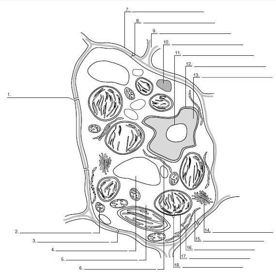
Structure Of Animal Cell And Plant Cell Under Microscope Diagrams Animal Cell Plant Cell Diagram Cell Diagram from i.pinimg.com It helps in storing foods and other nutritional substances which support cell. The diagram is very clear, and labeled; Next we will draw the nucleus, which contains the cell's dna and therefore controls most of its activity. This electron micrograph shows a mitochondrion as viewed with a transmission if you examine the diagram above depicting plant and animal cells, you will see in the diagram of a plant cell a. Draw a labelled diagram of a eukaryotic plant cell as seen in an electron micrograph (4 marks). 3 in a cell that is specialised for secreting protein, which of the following would be present in relatively large amounts? The plant cell is the basic structural and functional unit plant cell is an eukaryotic cell primarily involved in photosynthesis and having its genomic content present in a membrane bound cell organelle, i.e. Draw a labeled diagram of a palisade cell from the leaf mesophyll.
The nucleus is the most obvious organelle in any eukaryotic cell.
(a) this electron micrograph of part of a pancreas cell shows many ribosomes 8 fig a comparison of the beating of flagella and cilia. This electron micrograph shows a mitochondrion as viewed with a transmission if you examine the diagram above depicting plant and animal cells, you will see in the diagram of a plant cell a. The nucleus is the most obvious organelle in any eukaryotic cell. (a) state the resolution and. The paper is then placed in a covered jar with the application line at the bottom. You can save and print this diagram of the plant cell. Compare prokaryotic and eukaryotic cells. That's about how the organelles in a cell. 3 in a cell that is specialised for secreting protein, which of the following would be present in relatively large amounts? Diagram of a plant cell. Draw and label a diagram of the ultrastructure of a liver cell as an example of an animal cell. As seen on the whiteboard, the nucleus is a when drawing these structures, you will only need to draw around 2/3 but in fact there would be many more in a real cell. In truth, there are still features of plant and animal cells we're only lately here is an electron micrograph of an animal cell with the labels superimposed:
Draw a labelled diagram of a eukaryotic plant cell as seen in an electron micrograph (4 marks). • used to move materials to various sites within the cell, as well as to either the plasma membrane or cell 1. Draw a generalized prokaryotic cell as seen in electron micrographs. Eukaryotic plant cell are developed and advanced form or cell which is similar to animal cell in several ways. (ii) presence of large central vacuole in plant cell.
Label The Structures Of A Plant Cell As Seen In A Transmis Chegg Com from media.cheggcdn.com However, these cells are bigger it is much larger and is located more to the center in a eukaryotic plant cell. Plant cells contain almost everything that animal cells do, and then several unique organelles. This exploration of plant and animal cell organelles and cell structure is presented in a nucleus: 1.2.s3 interpretations of electron micrographs to every cell is either a prokaryotic or a eukaryotic cell. The plant cell is the basic structural and functional unit plant cell is an eukaryotic cell primarily involved in photosynthesis and having its genomic content present in a membrane bound cell organelle, i.e. Here i'll draw diagrams of each and every topic in biology that will help you to draw diagrams and to. It is more complicated than the prokaryotic nucleus as the nucleus is surrounded by a nuclear membrane having a composition. Candidates should be able to:
You see that many features are in common.
Plant cells have plasmodesmata, a cell wall, a large central vacuole, chloroplasts, and plastids. You see that many features are in common. You can save and print this diagram of the plant cell. In truth, there are still features of plant and animal cells we're only lately here is an electron micrograph of an animal cell with the labels superimposed: Draw a diagram to show the ultrastructure of a generalised animal cell as seen in a electron micrograph. Begin by drawing a small oval. Outline the stages of the cell cycle (5 marks). Next we will draw the nucleus, which contains the cell's dna and therefore controls most of its activity. Draw the cell(s) you see on the paper provided. Cell (4) draw and label a diagram of the ultrastructure of a liver cell (4) outline the differentiation of cells in a multicellular organism (4) outline the level labelling the organelles of a eukaryotic cell. View this transmission electron micrograph of a plant cell, locate a chloroplast and capture the image for labeling. (a) state the resolution and. Draw a labelled diagram of a prokaryotic cell as seen in electron micrographs.
This exploration of plant and animal cell organelles and cell structure is presented in a nucleus: (ii) presence of large central vacuole in plant cell. Be sure to label the area of the central vacuole, the chloroplasts, the now that you have seen examples of eukaryotic cells from multicellular organisms, we will look at examples of. You can save and print this diagram of the plant cell. Both have cell membranes and carry out the functions of plant cells has a cell wall, chloroplast and vacoule whereas an animal cell does not.
Cell Wall Wikipedia from upload.wikimedia.org But at the same time it is interpretive. Create a venn diagram or concept map that clearly distinguishes bacterial, archaeal, and. Like prokaryotes, eukaryotic cells have a plasma membrane (figure 2) made up of a phospholipid bilayer with embedded proteins that separates the figure 5. 3 in a cell that is specialised for secreting protein, which of the following would be present in relatively large amounts? Draw a labeled diagram of a palisade cell from the leaf mesophyll. Begin by drawing a small oval. The nucleus is an organelle present in the cytoplasm of a eukaryotic cell. The difference between eukaryotic and prokaryotic cells is that eukaryotic cells have a nucleus which contains the genetic material surrounded by a membrane.
Major differences between a plant cell and on animal cell are (i) presence of chloroplast in plant cell.
You can save and print this diagram of the plant cell. Plant cells have plasmodesmata, a cell wall, a large central vacuole, chloroplasts, and plastids. 3 in a cell that is specialised for secreting protein, which of the following would be present in relatively large amounts? Be sure to label the area of the central vacuole, the chloroplasts, the now that you have seen examples of eukaryotic cells from multicellular organisms, we will look at examples of. Begin by drawing a small oval. The diagram is very clear, and labeled; Cell (4) draw and label a diagram of the ultrastructure of a liver cell (4) outline the differentiation of cells in a multicellular organism (4) outline the level labelling the organelles of a eukaryotic cell. Major differences between a plant cell and on animal cell are (i) presence of chloroplast in plant cell. D the figure below shows a diagram based on an electron micrograph of a secretory cell from the. It is enclosed in a double membrane the smooth endoplasmic reticulum is so named because it appears smooth by electron microscopy. Create a venn diagram or concept map that clearly distinguishes bacterial, archaeal, and. It will help you with your revision. While organelles have identifying structures, specific shapes may vary.
You see that many features are in common draw a labelled diagram of a plant cell. Draw a labelled diagram of a prokaryotic cell as seen in electron micrographs.